Dental photography
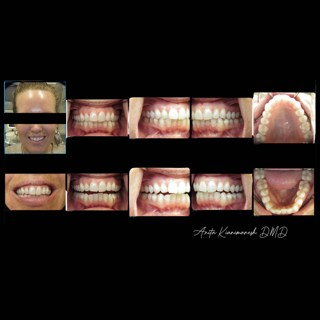
Intra & Extra-Oral Photos
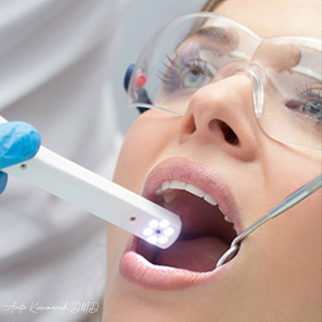
Achieve a clearer picture of your oral health with our advanced intraoral photography technology. High-resolution images allow your dentist to provide a more comprehensive evaluation, leading to a personalized treatment plan for a healthier smile.
See More, Smile More: Intraoral Imaging for Optimal Care
Intraoral photos provide detailed views of your teeth and gums, revealing cracks, decay, gum disease, and other potential issues. These magnified images can help your dentist diagnose problems early, monitor treatment progress, and explain treatment options more effectively.


What Are Intra-Oral Photos?
Intraoral photography is a valuable dental diagnostic tool that utilizes a small, high-resolution camera to capture close-up images of the inside of your mouth. These detailed pictures provide dentists with a magnified view of your teeth, gums, tongue, and other oral tissues, allowing them to identify a wide range of dental concerns.
Intraoral photos can be instrumental in detecting cavities, cracks, gum disease (gingivitis and periodontitis), tooth wear, misalignment, and even cancerous lesions in their early stages.
They can also be used to assess the health and position of implants, dentures, and other dental restorations.
Frequently Asked Questions
Yes, intraoral photography is a safe and painless procedure. The specialized cameras emit minimal light and do not generate harmful radiation.
These close-up images can reveal subtle details invisible to the naked eye, allowing for earlier diagnosis and more accurate treatment plans.
The intraoral camera is small and flexible, causing minimal discomfort. Most patients experience no pain during the process.
Intraoral photos become part of your dental records. They are used for diagnosis, treatment planning, tracking progress, and insurance purposes.

